We use cookies to ensure that we give you the best experience on our website. If you continue to use this site, you consent to the use of these cookies. OK

For US Healthcare Professionals Only

Resources

Videos

Understanding CTCL

Mara Haseltine, MD

Understanding CTCL

[Presenter: Mara Haseltine, MD, Dermatologist and Director of the University Medical Center Cutaneous T Cell Lymphoma Clinic. Mara Haseltine, MD was compensated by Kyowa Kirin for her participation in this video.]

[VISUAL ON SCREEN: Pie chart highlighting the CBCL/CTCL/Mycosis Fungoides/Sézary Syndrome sections as Dr. Haseltine speaks to each.]

DR. HASELTINE: Cutaneous T-cell lymphomas, or CTCL, are a type of non-Hodgkin lymphoma. They account for the vast majority of primary cutaneous lymphomas.1 The CTCL subtype Mycosis Fungoides, or MF, is more common—comprising around 60 percent of CTCL cases.1 Sézary Syndrome, or SS, is more rare; as you can see, it makes up roughly 5%.2 Together, Mycosis Fungoides and Sézary Syndrome comprise around two-thirds of all CTCL cases.1

CTCL primarily manifests in the skin, but other disease compartments may also be involved, including lymph nodes, viscera, and blood.1,3 Mycosis Fungoides may be indolent, with skin-only symptoms. Still, it has a heterogeneous presentation even within skin.4,5

Mycosis Fungoides may also involve other disease compartments.6,7 In fact, 1 in 3 patients with Mycosis Fungoides may experience progression within skin It is associated with poorer prognosis.or to other compartments, including blood.7

Sézary Syndrome is a more aggressive subtype.4 It is defined by high levels of blood involvement and erythroderma, intense pruritus, and often lymphadenopathy.1 It is associated with poorer prognosis.7

Blood classification and disease stage

DR. HASELTINE: Now, let’s take a closer look at how Mycosis Fungoides and Sézary Syndrome are staged, and how staging can inform prognosis.

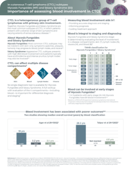
[VISUAL ON SCREEN: Table showing TNMB criteria for skin, lymph node, viscera, and blood involvement.]

DR. HASELTINE: Clinical stage is determined using the TNMB system. It evaluates the level of involvement in each of the disease compartments.8 In the TNMB classification system:

  • T represents the extent of skin involvement, as well as lesion types
  • Lymph node, or N, is determined by histopathological features of lymph node abnormalities
  • Viscera, or M, depicts the absence or presence of metastatic involvement
  • Blood, or B, highlights the level of blood involvement in the disease8

Blood classification was added to the TNM criteria for Mycosis Fungoides and Sézary Syndrome in 2007 as an independent prognostic factor.9

[VISUAL ON SCREEN: Table showing NCCN blood classifications for Mycosis Fungoides and Sézary Syndrome.]

DR. HASELTINE: Blood involvement is determined by counts of abnormal cells and T-cell clone status, and is classified as8:

  • B0—no significant blood involvement
  • B1—low blood involvement
  • B2—high blood involvement

[VISUAL ON SCREEN: Table showing blood classifications.]

DR. HASELTINE: Here are the full classifications for B0 to B2 blood involvement. To review them, feel free to pause the video, or explore www.PROBEinCTCL.com.

Clinical staging

[VISUAL ON SCREEN: Table showing TNMB classifications for Mycosis Fungoides and Sézary Syndrome. The Clinical Stage column is highlighted first, then the TNMB columns are highlighted.]

DR. HASELTINE: Next, this is how clinical stage is determined, based on combining all four TNMB criteria, and the TNMB classifications that comprise each clinical stage are shown here.8

[VISUAL ON SCREEN: Table showing TNMB classifications for Mycosis Fungoides and Sézary Syndrome. The Stage IVA1, IVA2, and IVB rows are highlighted, then the B column is highlighted.]

DR. HASELTINE: While high blood involvement is always considered advanced disease, regardless of other compartmental involvement,8 blood can be involved even in early-stage disease.6

[TEXT ON SCREEN: 1 in 5 early-stage Mycosis Fungoides patients have blood involvement. FOOTNOTE: Based on a PROCLIPI database study that analyzed hematological data from 348 patients with early-stage Mycosis Fungoides6]

DR. HASELTINE: In fact, one study found that roughly 1 in 5 patients with early-stage Mycosis Fungoides have B1 blood involvement.6

[VISUAL ON SCREEN: Chart showing median overall survival by blood classification.]

DR. HASELTINE: But why is blood involvement so important?

In Mycosis Fungoides and Sézary Syndrome, blood is an independent prognostic factor,9 and is associated with worse outcomes.7 Shorter overall and disease-specific survival have been correlated with increasing blood involvement.7 In fact, one study showed that patients with B2, or high blood involvement, have a 4.6x greater risk of disease progression.7

Now that I’ve discussed the importance of blood involvement in Mycosis Fungoides and Sézary Syndrome, what does the workup look like?

No single diagnostic test is available for Mycosis Fungoides or Sézary Syndrome. Because these are multicompartmental diseases, NCCN guidelines recommend a multimodal approach for diagnosing and staging.8 A full workup with evaluations of the 4 compartments is important for accurate diagnosis and staging. Workup should include8:

  • Skin exam and biopsy
  • Palpation and CT scan of lymph nodes
  • Palpation and CT scan of viscera
  • Flow cytometry to assess blood involvement8

Flow cytometry for blood involvement

DR. HASELTINE: Flow cytometry is an assay that is ordered from the lab. It provides qualitative and quantitative measures of abnormal T-cells in blood.10,11 Flow cytometry should be done at diagnosis, and when certain clinical flags appear throughout the course of your patient’s journey with Mycosis Fungoides and Sézary Syndrome.8,12-14

To learn more about when to order flow cytometry, and how flow cytometry works, watch the video in this series presented by Dr. Alejandro Gru.

Here is the key takeaway from this video: Blood involvement is an important prognostic factor, and it should be monitored both in Mycosis Fungoides and Sézary Syndrome patients.

I hope the information in this video is useful. Thank you for watching. For more information about flow cytometry for blood involvement, as well as example clinical cases, explore www.PROBEinCTCL.com.

References: 1. Willemeze R, et al. Blood. 2019;133:1703-1714. 2. National Institute of Health - SEER Program - Sezary syndrome. Accessed September 19, 2022. https://seer.cancer.gov/seertools/hemelymph/51f6cf56e3e27c3994bd52e2/. 3. Olsen E, et al. Blood. 2022;140(5):419-437. 4. Willemze R, et al. Blood. 2005;105:3768-3785. 5. Cerroni L. Semin Cutan Med Surg; 2018;37:2-10. 6. Scarisbrick JJ et al. Br J Dermatol. 2019;181:350-357. 7. Agar NS, et al. J Clin Oncol. 2010;28(31):4730-4739. 8. NCCN Guidelines. Primary cutaneous lymphomas (v.2.2022). www.nccn.org. Accessed November 1, 2022. 9. Olsen E, et al. Blood. 2007;110:1713-172. 10. Illingworth A, et al. Cytometry B Clin Cytom. 2021;100(2):156-182. 11. Craig FE. Cytometry. 2021;100:125-128. 12. Vermeer MH, et al. Br J Dermatol. 2022;187:21-28. 13. Scarisbrick JJ, et al. Eur J Cancer. 2018;93:47-56. 14. Willemze R, et al. Ann Oncol. 2018;29 (suppl4):iv30-iv40.

Diagnosing blood involvement in CTCL

Alejandro A. Gru, MD

Diagnosing blood involvement in CTCL

[Presenter: Alejandro A. Gru, MD, Dermatopathology Fellowship Director, University of Virginia. Alejandro Gru, MD was compensated by Kyowa Kirin for his participation in this video.]

DR. GRU: CTCL subtypes Mycosis Fungoides, or MF, and Sézary Syndrome, or SS, are multicompartmental diseases.1 That’s why NCCN Guidelines recommend a multimodal diagnostic approach for optimal patient management.2 As we’ll discuss, this includes using flow cytometry for evaluating blood involvement when appropriate.2

To learn more about why blood involvement matters in Mycosis Fungoides and Sézary Syndrome, watch the video in this series presented by Dr. Mara Haseltine, Dermatologist and Director of the University Medical Center Cutaneous T Cell Lymphoma Clinic.

Flow cytometry for blood involvement

DR. GRU: Flow cytometry measures blood involvement in Mycosis Fungoides and Sézary Syndrome.1 It provides qualitative and quantitative measures of abnormal T-cells, or Sézary cells, in blood.3

[VISUAL ON SCREEN: Diagram of cells passing through a flow cytometer, producing multicolor light scatter as they pass through a laser.]

DR. GRU: The advantages of flow cytometry are, it is:

  • Robust4
  • Quantifiable5
  • Objective5
  • Consistent6
  • Technologically precise7,8
  • Becoming more widely used versus manual counts5

For all these reasons, flow cytometry is recommended in US and international Guidelines (NCCN, ISCL, USCLC, EORTC) for evaluating blood involvement in CTCL.2,5

So, how does flow cytometry work?

[VISUAL ON SCREEN: Diagram of a beaker with cells suspended in solution.]

DR. GRU: First, cells are tagged with fluorescent markers of interest and suspended in solution.

[VISUAL ON SCREEN: Diagram of cells passing through a flow cytometer, producing light scatter as they pass through a laser.]

These fluorescent cells are then injected into the flow cytometer, and are evaluated as they flow past multiple lasers. Each cell is analyzed for visible light scatter and multiple fluorescence parameters.10

[VISUAL ON SCREEN: Diagram of a computer.]

DR. GRU: This light scatter information is collected and processed by a computer.10

[VISUAL ON SCREEN: Diagram of a report form.]

DR. GRU: Then, the computer-generated output provides the basis for diagnosis. The report contains data on abnormal T-cell populations.3

[VISUAL ON SCREEN: Chart of T-cell panel markers CD3, CD4, CD8, CD45, CD7, and CD26.]

DR. GRU: No single T-cell marker can accurately identify Sézary cells by itself. To diagnose Mycosis Fungoides or Sézary Syndrome, Guidelines developed by an international group of cytometry specialists recommend a minimum of 6 panel markers.11

The panel markers that are used may vary between flow cytometry labs. At a minimum, they should include these 6, but labs may use additional markers as well.11 These markers help identify and quantify T-cells and detect Sezary cells.11

[VISUAL ON SCREEN: Chart of T-cell panel markers CD7 and CD26.]

DR. GRU: The most common T-cell abnormalities seen in Mycosis Fungoides and Sézary Syndrome are loss of CD7 and/or CD26.5 In fact, loss of CD7 has been identified in 50% to 80% of cases with blood involvement, and loss of CD26 has been identified in 70% to 100% of cases with blood involvement.5

When requesting flow cytometry, a complete blood count should also be ordered to help calculate absolute numbers of abnormal cells.8,11

The flow cytometry report

[VISUAL ON SCREEN: Example report showing the inclusion of negative/positive diagnosis and clinical interpretation of results.]

DR. GRU: Although flow cytometry reports can vary between labs, the report you receive should provide a clinical interpretation of the results.8, 11 This statement will report the presence or absence of abnormal T cells, and whether the results are consistent with CTCL.3

[VISUAL ON SCREEN: Example report showing quantitative T-cell panel results, including absolute counts and percentages.]

DR. GRU: The report typically includes quantitative T-cell panel results, including absolute counts of T-cells and percent of total lymphocytes, as well as CD4:CD8 T-cell ratios.3,8 Blood classification may be specified.

Flow cytometry guidelines

DR. GRU: So, when might you consider flow cytometry? European Guidelines (ESMO, EORTC) recommend flow cytometry for measuring blood involvement at all stages of Mycosis Fungoides and Sézary Syndrome.12,13

NCCN Guidelines state that flow cytometry may be useful at diagnosis. Additionally, NCCN recommends that flow cytometry be included in the workup for any patients with T2-T4 skin classification, and any suspected extracutaneous disease.2

According to recently published consensus guidance, flow cytometry is recommended: at diagnosis, when certain clinical flags appear, and during follow-up.5,6

Here are the consensus recommendations. At diagnosis, an initial blood evaluation for all suspected Mycosis Fungoides or Sézary Syndrome can help with:

  • Accurate diagnosis, staging, and informing prognosis
  • Establishing a baseline, which is important for monitoring changes in blood involvement over time
  • Guiding treatment approach5

Flow cytometry is also recommended when certain clinical flags appear, such as in5:

  • Disease progression
  • Patients with advanced disease
  • Intractable pruritus
  • Generalized patches and/or plaques
  • Erythroderma
  • Lymphadenopathy

Flow cytometry may also be considered in the case of abnormal lab results, such as5:

  • Lymphocytosis on white blood cell count
  • High serum LDH
  • Lack of response to treatment

Other flags may include lymphocytosis with hypercalcemia, or absolute eosinophilia.

How often might you consider flow cytometry during follow-up with your patient?5 Every 3 months is recommended where practical, or in the case of abnormal flow cytometry at baseline. Flow cytometry should also be considered if there is disease or stage progression, or upon the development of any clinical flags.

And if the flow cytometry results are abnormal, T-cell clonality studies should be done as follow-up to confirm clonality in diagnosis and monitoring.5 NCCN Guidelines recommend correlating TCR studies with molecular findings from skin to determine if clones in the blood are the same as the ones in the skin.2

Tips for requesting flow cytometry

DR. GRU: Lastly, when requesting flow cytometry, keep these things in mind to optimize utility of the test:

  • Specify assay objectives when ordering flow cytometry, such as “Rule out Mycosis Fungoides or Sézary Syndrome”
    • This is important for the pathologist to choose the appropriate flow cytometry panel.3 And remember, guidelines recommend a minimum of 6 T-cell markers11
  • Include clinical details
    • A detailed description of clinical history and observations, along with any existing diagnosis, will be helpful for the pathologist to interpret results11
  • Use the same flow cytometry lab routinely
    • This may help ensure consistency in the flow cytometry methodology and the report summary8,11
  • Consider consultation with a colleague who specializes in CTCL
    • A multidisciplinary team and consultation at a center with CTCL expertise is recommended in the NCCN Guidelines for optimal patient management2

Thank you for watching. I hope you found this video helpful. For more information about blood involvement in Mycosis Fungoides and Sézary syndrome, including example clinical cases, explore www.PROBEinCTCL.com

References: 1. Olsen EA, Whittaker S, Willemze R, et al. Primary cutaneous lymphoma: recommendations for clinical trial design and staging update from the ISCL, USCLC, and EORTC. Blood. 2022;140(5):419-437. 2. Referenced with permission from the NCCN Clinical Practice Guidelines in Oncology (NCCN Guidelines®) for Primary Cutaneous Lymphomas V.2.2022. © National Comprehensive Cancer Network, Inc. 2022. All rights reserved. Accessed October 1, 2022. 3. Illingworth A, Johansson U, Huang S, et al. International guidelines for the flow cytometric evaluation of peripheral blood for suspected Sézary syndrome or mycosis fungoides: assay development/optimization, validation, and ongoing quality monitors. Cytometry B Clin Cytom. 2021;100(2):156-182. 4. Tembhare PR, Chatterjee G, Chaturvedi C, et al. Critical role of flkow cytometric immunophenotyping in the diagnosis, subtyping, and staging of T/NK-cell non-Hodgkin’s lymphoma in real-world practice: A study of 232 cases from a tertiary cancer center in India. Front Onc. 2022;12:doi: 10.3389/fonc.2022.779230. 5. Vermeer MH, Moins-Teisserenc H, Bagot M, et al. Flow cytometry for the assessment of blood tumour burden in cutaneous T-cell lymphoma: towards a standardized approach. Br J Dermatol. 2022;187(1):21-28. 6. Vermeer MH, Nicolay JP, Scarisbrick JJ, Zinzani PL. The importance of assessing blood tumour burden in cutaneous T-cell lymphoma. Br J Dermatol. 2020;185(1):19-25. 7. Craig F, KA Foon. Flow cytometric immunophenotyping for hematologic neoplasms. Blood. 2008; 111 (8): 3941–3967. 8. Guitart J. Sézary syndrome and mycosis fungoides flow cytometric evaluation: the clinicians’ perspective. Cytometry B Clin Cytom. 2021;100 (2):129-131. 9. Vermeer MH, Nicolay JP, Scarisbrick JJ, Zinzani PL. The importance of assessing blood tumour burden in cutaneous T-cell lymphoma. Br J Dermatol. 2020;185(1):19-25. 10. McKinnon KA. Flow cytometry: an overview. Curr Protoc Immunol; 2018;120:5.1.1-5.1.11. 11. Horna P, et al. Flow cytometric evaluation of peripheral blood for suspected Sézary syndrome or mycosis fungoides: international guidelines for assay characteristics. Cytometry B Clin Cytom. 2021;100(2):142-155. 12. Scarisbrick JJ, Quaglino P, Prince HM, et al. The PROCLIPI international registry of early-stage mycosis fungoides identifies substantial diagnostic delay in most patients. Br J Dermatol. 2019;181:231-232. 13. Willemze R, Hodak E, Zinzani PL, et al. Primary cutaneous lymphomas: ESMO Clinical Practice Guidelines for diagnosis, treatment and follow-up. Ann Oncol. 2018 Oct 1;29(Suppl 4):iv30-iv40. doi: 10.1093/annonc/mdy133.

Commentary on the 5 example clinical cases

Psoriasis or Mycosis Fungoides?

Joan Guitart, MD

Patient Case 1

[Presenter: Joan Guitart, MD, Professor of Dermatology and Pathology, Northwestern University. All treatment decisions described in this video are the opinions of Dr. Guitart. Joan Guitart, MD was compensated by Kyowa Kirin for his participation in this video.]

DR. GUITART: Blood involvement in cutaneous T-cell lymphoma can occur even at early stages of Mycosis Fungoides and, by definition, in Sézary Syndrome.1

In this video series, I will discuss hypothetical example patient cases where flow cytometry was ordered to assess blood involvement. These examples may be similar to patients you see in your practice.

Let’s discuss Patient 1 to find out why flow cytometry was ordered.

[VISUAL ON SCREEN: Image of a 70-year-old man. Not a real patient. Image courtesy of Joan Guitart, MD.]

DR. GUITART: Patient 1 is a 70-year-old male, who has been treated for plaque psoriasis for 5 years. He has patches on his legs and arms, including elbows, and lower back, and he is also experiencing pruritus—this clinical presentation is entirely consistent with psoriasis.

[VISUAL ON SCREEN: Photo of skin symptoms. Image courtesy of Joan Guitart, MD.]

DR. GUITART: Since his diagnosis 5 years ago, his treatments have included topical steroids, phototherapy, and biologics. He demonstrated a partial response to each treatment, but experienced subsequent worsening of skin symptoms. Most recently, he was treated with a TNF-alpha inhibitor, but his skin symptoms persisted.

In my experience, Mycosis Fungoides does not typically present like this. As you can see in this image, patient presented with plaques that resemble psoriasis, with thick scale and marked erythema.

[VISUAL ON SCREEN: Photo of skin symptoms. Image courtesy of Joan Guitart, MD.]

DR. GUITART: He is currently presenting with worsening skin symptoms and increased pruritus. Patches and plaques are now more extensive, currently affecting close to 10% of his body skin surface, with more pronounced erythema.

This patient is failing standard therapy for psoriasis with worsening skin signs and symptoms. So, what would be my next steps?

Because the initial presentation was clinically consistent with psoriasis, he was never biopsied. At this point—2 to 3 weeks after topical therapy was discontinued1—I would choose to do a skin biopsy. And I always keep in mind that multiple biopsies of more than one lesion having different clinical appearances may be necessary to help reach a precise diagnosis in such a typical presentations.1

If involvement of hair follicles is suspected due to alopecia, a deeper punch biopsy may be preferable.1

His workup included complete blood count, metabolic panel and serum LDH, as well as palpation of lymph nodes, and punch skin biopsies.1 Complete blood count showed a lymphocyte count of 3100 lymphocytes/μL. His serum LDH levels and metabolic panel were normal, and adenopathy was not identified.

[VISUAL ON SCREEN: Image of punch biopsy. Image courtesy of Joan Guitart, MD.]

DR. GUITART: Punch biopsy of a representative skin lesion was reviewed by a dermatopathologist with expertise in cutaneous lymphomas. At low power, we appreciate marked epithelial hyperplasia with parakeratosis in a pattern resembling psoriasis.

[VISUAL ON SCREEN: Image of punch biopsy. Image courtesy of Joan Guitart, MD.]

DR. GUITART: But at higher power, the presence of abnormal medium sized lymphocytes is noted in the intraepidermal compartment.

Immunohistochemistry analysis revealed a predominance of CD4+ T-cells with deletion of CD7 and a high CD4:CD8 ratio of 10:1. The T-cell receptor gene rearrangement assay revealed a clonal population.1

So, why was flow cytometry ordered for this patient? What clinical flags would raise suspicion of Mycosis Fungoides and lead to further investigation?

We have a patient here with lesions resembling psoriasis, but refractory to standard psoriasis treatment. His skin symptoms were worsening despite intense skin-directed and systemic therapy, with more pruritus, and more extensive skin involvement. Now we have abnormal lab results, and the pathology report raises suspicion of Mycosis Fungoides.

So for next steps, I would want to assess the implications of lymphocytosis and order flow cytometry to assess for possible blood involvement.1,2

Flow cytometry results

[VISUAL ON SCREEN: Sample report showing flow cytometry results, not from an actual patient.]

DR. GUITART: Flow cytometry results showed no detectable blood involvement. Therefore, his negative blood assessment corresponds with a B0 blood classification, along with N0 due to lack of adenopathy indicating that this patient appears to have stage 1 MF with limited skin disease without extracutaneous involvement.3

The blood flow cytometry analysis shows 3100 lymphocytes/μL with an absolute T-cell count of 2300, with the remaining 800 representing B-cells and natural killer cells.

The total CD4 count is 1600 with a normal CD4:CD8 ratio of 2.3 and small percentage of cells without CD7 or CD26 expression. This distribution of T-cells is normal indicating lack of CTCL blood involvement.3

In summary, here are some clinical considerations related to this patient’s case:

  • Mycosis Fungoides can present with atypical signs
  • Mycosis Fungoides can mimic psoriasis, eczema, as well as other inflammatory skin conditions, as was the case with this patient8
  • While flow cytometry analysis was negative in this case, keep in mind that 1 in 5 patients with early Mycosis Fungoides may have blood involvement5

And given the rarity of the disease and the challenges with diagnosis, NCCN Guidelines recommend consultation with a specialist with CTCL expertise whenever Mycosis Fungoides or Sézary Syndrome is suspected—this was important for accurately diagnosing this patient.1

Also keep in mind recommendations from NCCN guidelines for when to consider flow cytometry:

  • NCCN Guidelines recommended flow cytometry for all patients with a new diagnosis of Mycosis Fungoides with body skin surface involvement of more than 10%, and for classification T3, which is the presence of tumors1
  • Based on the NCCN guidelines, I consider flow cytometry whenever Sézary Syndrome is suspected

Why should flow cytometry be considered? Because:

  • It helps with accurate staging, which provides prognostic information, and may guide treatment approach2
  • It also helps to establish baseline blood involvement, which can be monitored for changes over time2

When ordering flow cytometry keep in mind these considerations:

  • In my experience, it is important to specify a clinical suspicion for Mycosis Fungoides or Sézary Syndrome. This will alert the pathologist to include CTCL-specific T-cell markers in the flow cytometry panel
  • The recommended T-cell panel should include: CD3, CD4, CD7, CD8, CD26, and CD459
  • Finally, when ordering flow cytometry, I’ve found that sending blood samples to the same laboratory may help ensure consistency in flow methodology and the report summary7

Thank you for watching, I hope this information is useful. For more information, including additional patient cases, explore www.PROBEinCTCL.com.

References: 1. Referenced with permission from the NCCN Clinical Practice Guidelines in Oncology (NCCN Guidelines®) for Primary Cutaneous Lymphomas V.1.2023. © National Comprehensive Cancer Network, Inc. 2023. All rights reserved. Accessed February 1, 2023. To view the most recent and complete version of the guideline, go online to NCCN.org. 2. Vermeer MH, Moins-Teisserenc H, Bagot M, et al. Flow cytometry for the assessment of blood tumour burden in cutaneous T-cell lymphoma: towards a standardized approach. Br J Dermatol. 2022;187(1):21-28. 3. Olsen EA, Whittaker S, Willemze R, et al. Primary cutaneous lymphoma: recommendations for clinical trial design and staging update from the ISCL, USCLC, and EORTC. 2022:140(5):419-437. 4. Merck Manual Professional Version website. https://www.merckmanuals.com/professional/hematology-and-oncology/leukopenias. Accessed February 27, 2023. 5. Cleveland Clinic Website. T-cells page. https://my.clevelandclinic.org/health/body/24630-t-cells. Accessed February 27, 2023. 6. University of Rochester Medical Center Website. CD4-CD8 Ratio. https://www.urmc.rochester.edu/encyclopedia/content.aspx?contenttypeid=167&contentid=cd4_cd8_ratio#:~:text=A%20normal%20CD4%2FCD8%20ratio,HIV. Accessed February 27, 2023. 7. Guitart J. Sézary syndrome and mycosis fungoides flow cytometric evaluation: the clinicians’ perspective. Cytometry B Clin Cytom. 2021;100 (2):129-131. 8. Scarisbrick JJ, Quaglino P, Prince HM, et al. The PROCLIPI international registry of early-stage mycosis fungoides identifies substantial diagnostic delay in most patients. Br J Dermatol. 2019;181:231-232. 9. Horna P, et al. Flow cytometric evaluation of peripheral blood for suspected Sézary syndrome or mycosis fungoides: international guidelines for assay characteristics. Cytometry B Clin Cytom. 2021;100(2):142-155.

Worsening skin symptoms

Joan Guitart, MD

Patient Case 2

[Presenter: Joan Guitart, MD, Professor of Dermatology and Pathology, Northwestern University. All treatment decisions described in this video are the opinions of Dr. Guitart. Joan Guitart, MD was compensated by Kyowa Kirin for his participation in this video.]

DR. GUITART: Blood involvement in cutaneous T-cell lymphoma can occur even at early stages of Mycosis Fungoides and, by definition, in Sézary Syndrome.1

In this video series, I will discuss hypothetical example patient cases where flow cytometry was ordered to assess blood involvement. These examples may be similar to patients you see in your practice.

Let’s discuss Patient 2 to find out why flow cytometry was ordered.

[VISUAL ON SCREEN: 40-year-old woman. Not a real patient.]

DR. GUITART: Patient 2 is a 40-year-old woman. She was initially diagnosed 2 years ago with stage 1B Mycosis Fungoides. Skin was the only involved compartment, classified as T2, involving approximately 12 percent of body skin surface. She had no lymphadenopathy, and flow cytometry utilizing a T-cell panel showed no evidence of blood involvement at diagnosis. Furthermore, there was no evidence of extracutaneous involvement by imaging. Patient 2 was treated with phototherapy and bexarotene, and her skin symptoms were well controlled for around 1 year.

[VISUAL ON SCREEN: Photo of skin symptoms. Image courtesy of Joan Guitart, MD.]

DR. GUITART: Currently, she is presenting with worsening skin symptoms, including an increasing extent of patches and plaques, now covering over 25% of body surface area. She has also recently developed multiple skin tumors, suggestive of disease progression.1

Let’s go over her workup and clinical findings1:

  • A complete blood cell count showed normal white blood cell distribution without lymphocytosis
  • Palpation showed enlarged axillary lymph nodes
  • PET/CT imaging revealed enlarged lymph nodes ranging from 2-3 cm with increased SUV uptake
  • Lymph node biopsies revealed dermatopathic changes with small clusters of abnormal medium sized lymphocytes

All of these are indicative of disease progression.

[VISUAL ON SCREEN: Image of skin biopsy. Image courtesy of Joan Guitart, MD.]

DR. GUITART: At this point, I would choose to perform new skin biopsies because of the abnormal lymph node findings along with the presence of new tumor lesions.

Skin biopsy of a tumor lesion showed a diffuse infiltrate composed of large cells, indicative of large-cell transformation.

[VISUAL ON SCREEN: Image of skin biopsy. Image courtesy of Joan Guitart, MD.]

DR. GUITART: Here, the large cells can be clearly seen in this high-power image. Now, before we move on, what clinical flags would lead me to suspect blood involvement and prompt a flow cytometry test?

In this case, clinical flags that raised my suspicion of disease progression including2:

  • Worsening skin symptoms with increased extent of affected skin
  • Changes in lesion type from plaque to tumor
  • Evidence of large-cell transformation on skin biopsy indicating disease acceleration
  • Lymphadenopathy was also observed, suggestive of early extracutaneous disease

In this patient, to me, these are all indicative of disease progression, and prompted further investigation, including1:

  • Biopsy of new skin tumors
  • Lymph node biopsy
  • Imaging
  • Repeat flow cytometry test

[VISUAL ON SCREEN: Sample report showing flow cytometry results, not from an actual patient.]

DR. GUITART: In this patient’s case, a blood sample was taken for flow cytometry. The blood sample, together with the skin biopsies were sent to the same lab to test for clonality, and to evaluate whether there were matching T-cell clones in the blood and skin.3

Flow cytometry showed a normal absolute lymphocyte count with a slight increase in the CD4:CD8 ratio of 5.0.4 The pathologist reported an abnormal discrete clone of CD4+/CD7-/CD26- abnormal cells accounting for 29 % of total lymphocytes or an absolute count of 850 cells/μL. This represents a B1 classification bordering on B2.1 Clonality was also confirmed by performing T-cell gene rearrangement analysis of the skin and blood demonstrating the same clonal signature at both sites.3

For consistency, I would send the blood samples to the same flow cytometry lab that made the initial diagnosis, to minimize variability due to differing methodologies and antibody panels across centers.

Because this patient progressed to a higher stage, in my experience, it’s important that she continue to be routinely monitored with flow cytometry to check for further signs of progression.2

In closing, keep in mind these clinical considerations and recommendations related to this patient’s case:

  • 1 in 3 patients with Mycosis Fungoides progress to later stage disease, which can affect skin, lymph nodes, blood, or other systemic compartments2.
  • Patients with abnormal flow and abnormal clones must be closely followed, because they may indicate evolving Sézary Syndrome3.

Per NCCN Guidelines, patients with disease progression ideally should be managed by a multidisciplinary team including oncologists, dermatologists, and pathologists with CTCL expertise.1

Thank you for watching, I hope this information is useful. For more information, including additional patient cases, explore PROBEinCTCL.com.

References: 1. Referenced with permission from the NCCN Clinical Practice Guidelines in Oncology (NCCN Guidelines®) for Primary Cutaneous Lymphomas V.1.2023. © National Comprehensive Cancer Network, Inc. 2023. All rights reserved. Accessed February 1, 2023. To view the most recent and complete version of the guideline, go online to NCCN.org. 2. Vermeer MH, Moins-Teisserenc H, Bagot M, et al. Flow cytometry for the assessment of blood tumour burden in cutaneous T-cell lymphoma: towards a standardized approach. Br J Dermatol. 2022;187(1):21-28. 3. Olsen EA, Whittaker S, Willemze R, et al. Primary cutaneous lymphoma: recommendations for clinical trial design and staging update from the ISCL, USCLC, and EORTC. Blood. 2022:140(5):419-437. 4. University of Rochester Medical Center Website. CD4-CD8 Ratio. https://www.urmc.rochester.edu/encyclopedia/content.aspx?contenttypeid=167&contentid=cd4_cd8_ratio#:~:text=A%20normal%20CD4%2FCD8%20ratio,HIV. Accessed February 27, 2023. 5. Merck Manual Professional Version website. https://www.merckmanuals.com/professional/hematology-and-oncology/leukopenias. Accessed February 27, 2023. 6. Cleveland Clinic Website. T-cells page. https://my.clevelandclinic.org/health/body/24630-t-cells. Accessed February 27, 2023.

Erythrodermic Mycosis Fungoides

Joan Guitart, MD

Patient Case 3

[Presenter: Joan Guitart, MD, Professor of Dermatology and Pathology, Northwestern University. Joan Guitart, MD was compensated by Kyowa Kirin for his participation in this video.]

[All treatment decisions described in this video are the opinions of Dr. Guitart.]

DR. GUITART: Blood involvement in cutaneous T-cell lymphoma can occur even at early stages of Mycosis Fungoides and by definition in Sézary Syndrome.1

In this video series, I will discuss hypothetical example patient cases where flow cytometry was ordered to assess blood involvement. These examples may be similar to patients you see in your practice.

Let’s discuss Patient 3 to find out why flow cytometry was ordered.

[TEXT ON SCREEN: Patient Case: Erythrodermic Mycosis Fungoides]

[VISUAL ON SCREEN: Photo of a man with short gray hair. TEXT ON SCREEN: Diagnosed 4 years ago with stage IIIB erythrodermic Mycosis Fungoides; low-level blood involvement (T4, N0, M0, B11 FOOTNOTE: Not a real patient]

DR. GUITART: Patient 3 is a 60-year-old man, who was initially diagnosed 4 years ago with stage 3B erythrodermic Mycosis Fungoides. At diagnosis he presented with skin stage T4, defined as diffuse erythroderma over 80% of body surface area. There was also no evidence of nodal or visceral disease.1 Flow cytometry utilizing a T-cell panel revealed a low level of abnormal T-cells, consistent with B1 blood involvement.1

[TEXT ON SCREEN: Treatment history: Extracorporeal photophoresis; pegylated interferon; partial response ~12 months, waning efficacy]

DR. GUITART: His most recent treatment was extracorporeal photopheresis and injections of pegylated interferon;1 partial response in skin was maintained for roughly 12 months, but now the efficacy is waning with recurrent erythroderma and pruritus.

[VISUAL ON SCREEN: Photo of a lower back with erythroderma. TEXT ON SCREEN: Erythrodermic Mycosis Fungoides and a history of abnormal T-cells in the blood and worsening pruritus. FOOTNOTE: Image courtesy of Joan Guitart, MD.]

DR. GUITART: While this patient did not fulfill diagnostic criteria for Sézary syndrome at diagnosis1, in my experience, relapsing erythroderma in patients with a history of blood involvement may indicate that his disease is evolving from erythrodermic Mycosis Fungoides to the leukemic CTCL variant, Sézary syndrome. Worsening erythroderma and pruritus may be a warning sign of increased risk of disease progression.2-4

[VISUAL ON SCREEN: Photo of hands showing skin symptoms. TEXT ON SCREEN: Acral fissuring and hyperkeratosis. FOOTNOTE: Image courtesy of Joan Guitart, MD.]

DR. GUITART: Currently, the patient is experiencing worsening skin symptoms, intense and intractable pruritus5 and erythroderma, with acral fissuring and hyperkeratosis of his hands and feet. Mild palpable lymphadenopathy was also noted, and he reports weight loss with persistent fatigue and malaise.

[VISUAL ON SCREEN: Table showing recommended T-cell panel for CTCL6]

DR. GUITART: All these signs and symptoms raise suspicion of disease progression, and would prompt further investigation, including a full blood workup including complete blood count with differential, complete metabolic panel, LDH and repeat flow cytometry analysis using a T-cell panel that includes the 6 recommended T-cell markers.1,6

Including CD4 and CD8 markers is important for determining T-cell subsets. This will also help determine the CD4:CD8 ratio, which can provide additional relevant information.6

[TEXT ON SCREEN: Clinical flags for flow cytometry4: Symptoms and/or signs of disease progression; Intractable pruritus; Worsening erythroderma; Lymphadenopathy]

DR. GUITART: So, what are the clinical flags that would lead me to refer this patient for repeat blood flow cytometry?4 In this case, the patient had signs of disease progression, including intractable pruritus, worsening erythroderma, and lymphadenopathy; his constitutional symptoms, specifically weight loss, fatigue and malaise, were also concerning to me.

[TEXT ON SCREEN: Refractory to current treatment]

DR. GUITART: This patient was refractory to current treatment.

[TEXT ON SCREEN: Abnormal blood flow results at diagnosis]

DR. GUITART: In my experience, the initial detection of an abnormal blood flow result at diagnosis was also an important consideration.

For consistency, I would send the blood samples to the same flow cytometry lab that performed the previous analysis in order to minimize potential variability due to different methodologies across centers.6,7

[VISUAL ON SCREEN: Table comparing flow cytometry results from the previous year to the current results]

DR. GUITART: Let’s take a look at the flow cytometry results. Comparing results from the last report to the current one showed increased blood involvement, with approximately 3500 Sézary cells per microliter as indicated by the absolute cell counts of CD4-positive, CD7-negative and CD26-negative T cells. Additional abnormal parameters were lymphocytosis8 and a high CD4 to CD8 ratio, equal to or higher than 10 to 19

These findings indicate evolution to B2 blood classification, which is a defining criteria for Sézary syndrome.1

In my practice, this patient would be started on a new systemic therapy and a repeat monthly blood analysis including flow cytometry would be scheduled to assess his response to the treatment.

[TEXT ON SCREEN: Clinical considerations: Blood involvement is associated with poor outcomes10; Routine flow cytometry recommended for patients with abnormal flow at baseline4; Flow cytometry can help monitor response to treatment and can help guide treatment approach4]

DR. GUITART: In closing, keep in mind these clinical considerations and recommendations related to this patient’s case:

  • Blood involvement can develop at any stage of Mycosis Fungoides1 and can progress to the leukemic form of CTCL which has been associated with poor outcomes.10
  • Consensus guidance recommends routine evaluation of blood by flow cytometry, every 3 months if feasible, in patients who have abnormal flow cytometry results at baseline.4
  • In addition, it's important to routinely monitor blood by flow cytometry to help assess response to treatment and to help guide treatment approach.4

Thank you for watching, I hope this information is useful. For more information, including additional patient cases, explore PROBEinCTCL.com.

References: 1. Referenced with permission from the NCCN Clinical Practice Guidelines in Oncology (NCCN Guidelines®) for Primary Cutaneous Lymphomas V.1.2023. © National Comprehensive Cancer Network, Inc. 2023. All rights reserved. Accessed March 13, 2023. To view the most recent and complete version of the guideline, go online to NCCN.org. 2. Serrano L, Martinez-Escala ME, Zhou XA, Guitart J. Pruritus in cutaneous T-cell lymphoma and its management. Dermatol Clin. 2018:36:245-258. 3. Hoppe RT, Kim YH. Clinical manifestations, pathologic features, and diagnosis of mycosis fungoides. UpToDate. https://www.uptodate. com/contents/clinical-manifestations-pathologic-features-and-diagnosis-of-mycosis-fungoides. Updated September 23, 2022. Accessed October 24, 2022. 4. Vermeer MH, Moins-Teisserenc H, Bagot M, et al. Flow cytometry for the assessment of blood tumour burden in cutaneous T-cell lymphoma: towards a standardized approac. Br J Dermatol. 2022;187(1):21-28. 5. Serrano L, Martinez-Escala ME, Zhou XA, Guitart J. Pruritus in cutaneous T-cell lymphoma and its management. Dermatol Clin. 2018:36:245-258. 6. Horna P, et al. Flow cytometric evaluation of peripheral blood for suspected Sézary syndrome or mycosis fungoides: international guidelines for assay characteristics. Cytometry B Clin Cytom. 2021;100(2):142-155. 7. Guitart J. Sézary syndrome and mycosis fungoides flow cytometric evaluation: The clinicians’ perspective. Cytometry. 2021;100:129-131. 8. Mayo Clinic Website. 9. Olsen E, Vonderheid E, Pimpinelli N, et al. Revisions to the staging and classification of mycosis fungoides and Sézary syndrome: a proposal of the International Society for Cutaneous Lymphomas (ISCL) and the cutaneous lymphoma task force of the European Organization of Research and Treatment of Cancer (EORTC). Blood. 2007;110(6):1713-1722 10. Agar NS, Wedgeworth E, Crichton S, et al. Survival outcomes and prognostic factors in mycosis fungoides/Sézary syndrome: validation of the revised International Society for Cutaneous Lymphomas/European Organisation for Research and Treatment of Cancer staging proposal. J Clin Oncol. 2010;28(31):4730-4739.

Severe skin infection

Joan Guitart, MD

Patient Case 4

[Presenter: Joan Guitart, MD, Professor of Dermatology and Pathology, Northwestern University. Joan Guitart, MD was compensated by Kyowa Kirin for his participation in this video.]

[All treatment decisions described in this video are the opinions of Dr. Guitart.]

DR. GUITART: Blood involvement in cutaneous T-cell lymphoma can occur even at early stages of Mycosis Fungoides and, by definition, in Sézary Syndrome.1

In this video series, I will discuss hypothetical example patient cases where flow cytometry was ordered to assess blood involvement. These examples may be similar to patients you see in your practice.

Let’s discuss Patient 4 to find out why flow cytometry was ordered.

[TEXT ON SCREEN: Patient Case: Mycosis Fungoides with severe skin infection]

[VISUAL ON SCREEN: Photo of a woman with black hair. TEXT ON SCREEN: Diagnosed 5 years ago with stage IB MF (T2, N0, M0, B0)1 FOOTNOTE: Not a real patient.]

DR. GUITART: Patient 4 is a 50-year-old female. She was initially diagnosed 5 years ago with stage IB Mycosis Fungoides, with patch lesions affecting approximately 25% of body surface area. Flow cytometry showed no blood involvement at the time, and there was no other evidence of extracutaneous disease.

[TEXT ON SCREEN: Treatment history: Skin-directed therapies; Pegylated interferon; Histone deacetylase (HDAC) inhibitor]

DR. GUITART: After initial treatment with skin-directed therapies, she received pegylated interferon, and experienced an initial partial response, which subsequently waned. She was then switched to an HDAC inhibitor and again achieved a partial response.

[VISUAL ON SCREEN: Photo of a torso showing skin symptoms. TEXT ON SCREEN: Skin infection + worsening skin lesions FOOTNOTE: Image courtesy of Joan Guitart, MD.]

DR. GUITART: She recently developed worsening pruritus and a severe skin infection that required hospitalization and IV antibiotics. Since then, she has continued to experience worsening skin lesions, now affecting over 30% of body surface area with thick plaques with yellow serous exudate indicative of impetigo and ulcerated tumors, as well as palpable adenopathy.

[TEXT ON SCREEN: Clinical findings: Skin culture positive for staphylococcus aureus (staph) infection; CBC showed mild neutrophilia]

DR. GUITART: Skin and nasal swab cultures were positive for staphylococcus aureus, confirming a staph infection. Her complete blo od count showed neutrophilia without lymphocytosis.

[TEXT ON SCREEN: Considerations for next steps: Extensive patch/plaque-stage Mycosis Fungoides (IB); Repeated signs of infection; Recent hospitalization for staph infection]

DR. GUITART: Before we move on, let’s consider next steps:

This patient had extensive patch stage Mycosis Fungoides (stage IB) and has been showing repeated signs of infection with recent hospitalization for staph infection.

[TEXT ON SCREEN: Dr. Guitart would consider: Treatment for the staph infection; Lymph node biopsy if persistent lymphadenopathy; Flow cytometry using a T-cell panel]

DR. GUITART: In this case, I would consider treating with oral antibiotics, diluted bleach baths and intense topical therapy with emollients and steroid creams. If there is persistence of adenopathy, a lymph node biopsy may be considered.1

We would also check blood flow cytometry utilizing a T-cell panel to monitor for progression. For consistency, I would send the blood samples to the same flow cytometry lab that performed the previous analysis in order to minimize potential variability due to different methodologies across centers.2,3

[TEXT ON SCREEN: Clinical flags for flow cytometry4,5: Recurrent staph infections; Worsening patches and plaques, new tumors; Enlarged lymph nodes]

DR. GUITART: In this patient’s case, what are the clinical flags that would prompt further investigation, including flow cytometry?

The severe and recurring staph infections, which prompted concerns that the disease may be progressing and impacting immunity.4,6

The worsening patches, plaques, and now tumor lesions with enlarged lymph nodes, were all suggestive of disease progression.1,7

[TEXT ON SCREEN: Considerations and recommendations from experts: Impaired immunity and increased risk of skin infection can occur in Mycosis Fungoides or Sézary Syndrome6; Infection may be indicative of disease progression4; Flow cytometry should be performed when there are signs of disease progression5]

DR. GUITART: In closing, keep in mind these clinical considerations and recommendations from experts relevant to this patient.

  • Patients with Mycosis Fungoides or Sézary Syndrome may have impaired immunity and increased risk of infection.6
  • Skin infection may be indicative of disease progression.4 Clinical signs of infection and/or ulceration should be worked up with bacterial cultures.1
  • Prompt antibiotic treatment of infection is important in patients with CTCL, because in my experience, bacterial infections can aggravate CTCL.1
  • Flow cytometry should be performed when there are clinical signs of progression, such as recurring infections and worsening skin disease.8

Thank you for watching, I hope this information is useful. For more information, including additional patient cases, explore PROBEinCTCL.com.

References: 1. Referenced with permission from the NCCN Clinical Practice Guidelines in Oncology (NCCN Guidelines®) for Primary Cutaneous Lymphomas V.1.2023. © National Comprehensive Cancer Network, Inc. 2023. All rights reserved. Accessed April 14, 2023. To view the most recent and complete version of the guideline, go online to NCCN.org. 2. Horna P, et al. Flow cytometric evaluation of peripheral blood for suspected Sézary syndrome or mycosis fungoides: international guidelines for assay characteristics. Cytometry B Clin Cytom. 2021;100(2):142-155. 3. Guitart J. Sézary syndrome and mycosis fungoides flow cytometric evaluation: The clinicians’ perspective. Cytometry. 2021;100:129-131. 4. Talpur R, Bassett R, Duvic M. Prevalence and treatment of Staphylococcus aureus colonization in patients with mycosis fungoides and Sézary syndrome. Br J Dermatol. 2008;159:105-112. 5. Vermeer MH, Moins-Teisserenc H, Bagot M, et al. Flow cytometry for the assessment of blood tumour burden in cutaneous T-cell lymphoma: towards a standardized approach. Br J Dermatol. 2022;187(1):21-28. 6. Hoppe RT, Kim YH. Clinical manifestations, pathologic features, and diagnosis of mycosis fungoides. UpToDate. https://www.uptodate. com/contents/clinical-manifestations-pathologic-features-anddiagnosis-of-mycosis-fungoides. Updated September 23, 2022. Accessed October 24, 2022. 7. Serrano L, Martinez-Escala ME, Zhou XA, Guitart J. Pruritus in cutaneous T-cell lymphoma and its management. Dermatol Clin. 2018:36:245-258. 8. Talpur R, Singh L, Daulat S, et al. Long-term outcomes of 1,263 patients with mycosis fungoides and Sézary syndrome from 1982 to 2009. Clin Cancer Res. 2012;18(18):5051-5060. 11. Farabi B, Seminario-Vidal L, Jamgochian M, et al. Updated review on prognostic factors in mycosis fungoides and new skin lymphoma trials. J Cosmet Dermatol. 2021;21(7):2742-2748.

Early-stage Mycosis Fungoides

Joan Guitart, MD

Patient Case 5

[Presenter: Joan Guitart, MD, Professor of Dermatology and Pathology, Northwestern University. Joan Guitart, MD was compensated by Kyowa Kirin for his participation in this video.]

[All treatment decisions described in this video are the opinions of Dr. Guitart.]

DR. GUITART: Blood involvement in cutaneous T-cell lymphoma can occur even at early stages of Mycosis Fungoides and by definition in Sézary Syndrome.1

In this video series, I will discuss hypothetical example patient cases where flow cytometry was ordered to assess blood involvement. These examples may be similar to patients you see in your practice.

Let’s discuss Patient 5 to find out why flow cytometry was ordered.

[TEXT ON SCREEN: Patient Case: Limited, patch-stage disease]

[VISUAL ON SCREEN: Photo of a man with short gray hair. TEXT ON SCREEN: Diagnosed 5 years ago with IA MF (T1, N0, M0, B0)1 FOOTNOTE: Not a real patient.]

DR. GUITART: Patient 5 is a 50-year-old male. He was initially diagnosed with stage IA Mycosis Fungoides 5 years ago.

[VISUAL ON SCREEN: Photo showing skin symptoms. TEXT ON SCREEN: Limited, patch-stage disease. FOOTNOTE: Image courtesy of Joan Guitart, MD.]

DR. GUITART: At initial presentation, he had patch lesions affecting approximately 5% of body surface area. Lymph nodes were normal on palpation, and CBC was normal as well, with no signs of lymphocytosis.

[VISUAL ON SCREEN: Table showing recommended T-cell panel for CTCL2]

DR. GUITART: Because this was a new diagnosis of Mycosis Fungoides, a blood sample was sent to assess for blood involvement by flow cytometry analysis using a T-cell panel that included the 6 recommended T-cell markers.1,3

Including CD4 and CD8 markers is important for determining T-cell subsets. This will also help determine the CD4:CD8 ratio, which can provide additional relevant information.2

Flow cytometry did not identify an abnormal T-cell population. Based on the workup and negative flow result, he was treated with phototherapy and topical steroids, which worked well to control the symptoms. He was followed up once a year to assess for disease progression. Follow-up exams included a full workup and flow cytometry.3

[VISUAL ON SCREEN: Photo showing increased skin symptoms. TEXT ON SCREEN: Increase in extent of patches (~15% of BSA), flow showed B1 blood classification. FOOTNOTE: Image courtesy of Joan Guitart, MD.]

DR. GUITART: On his most recent visit, he presented with patches and plaques now covering approximately 15% of body surface area. Flow cytometry was repeated, which now showed the presence of blood involvement, correlating with B1 classification, or low-level blood involvement.1

[TEXT ON SCREEN: Clinical considerations: Disease progression in skin and blood prompted more frequent monitoring5; Routine flow cytometry is recommended for patients with abnormal flow cytometry results5; Sending blood samples to the same flow cytometry center can help minimize variability2,6]

DR. GUITART: The increase in extent of affected skin, along with the detection of an abnormal T-cell population in the blood cell phenotype signals to me, a need to monitor this patient more closely. This disease acceleration would warrant closer follow up, and I would increase the frequency of flow cytometry from once a year to once every 3-6 months.4

For consistency, I would continue to send the blood samples to the same flow cytometry lab that performed the previous analyses in order to minimize potential variability due to different methodologies across centers.2,6

[TEXT ON SCREEN: Considerations and recommendations from experts: Although often indolent, up to 1 in 3 patients with Mycosis Fungoides may experience disease progression6; Flow cytometry is recommended at diagnosis4; Flow cytometry should be performed when there are signs of disease progression5]

DR. GUITART: So, keep in mind that, on diagnosis of Mycosis Fungoides, both a complete blood count and flow cytometry can provide a more complete diagnostic picture. Mycosis Fungoides is often indolent, but up to a third of patients may experience progression within skin or to other disease compartments, including blood.6

Flow cytometry at diagnosis, as well as throughout treatment, is recommended to establish baseline, and to monitor disease progression, in order to help guide treatment approach.4

But upon signs of progression, flow should be performed and repeated routinely, every 3-6 months depending on the signs and symptoms and disease acceleration.5

Thank you for watching, I hope this information is useful. For more information, including additional patient cases, explore PROBEinCTCL.com.

References: 1. Referenced with permission from the NCCN Clinical Practice Guidelines in Oncology (NCCN Guidelines®) for Primary Cutaneous Lymphomas V.1.2023. © National Comprehensive Cancer Network, Inc. 2023. All rights reserved. Accessed April 14,2023. To view the most recent and complete version of the guideline, go online to NCCN.org. 2. Horna P, et al. Flow cytometric evaluation of peripheral blood for suspected Sézary syndrome or mycosis fungoides: international guidelines for assay characteristics. Cytometry B Clin Cytom. 2021;100(2):142-155. 3. Olsen 2007. 4. Olsen EA, Whittaker S, Willemze R, et al. Primary cutaneous lymphoma: recommendations for clinical trial design and staging update from the ISCL, USCLC, and EORTC. Blood. 2022;140(5):419-437 5. Vermeer MH, Moins-Teisserenc H, Bagot M, et al. Flow cytometry for the assessment of blood tumour burden in cutaneous T-cell lymphoma: towards a standardized approach. Br J Dermatol. 2022;187(1):21-28. 6. Guitart J. Sézary syndrome and mycosis fungoides flow cytometric evaluation: The clinicians’ perspective. Cytometry. 2021;100:129-131. 7. Agar NS, Wedgeworth E, Crichton S, et al. Survival outcomes and prognostic factors in mycosis fungoides/Sézary syndrome: validation of the revised International Society for Cutaneous Lymphomas/European Organisation for Research and Treatment of Cancer staging proposal. J Clin Oncol. 2010;28(31):4730-4739.

Downloadable materials